FeMO2 Dive Cruise 2007
Report -- Deep Sea Bacteria Spin Rust and Eat Nails for a Living
FeMO2 Dive Cruise 2007 |
Spinning Rust
The microbial mats at Loihi can be thought of as a giant fabric woven together by the microorganisms that grow there. It is the bacteria that are responsible for spinning the iron oxides into the filaments or threads that create the larger mat fabric. To the eye the mats appear to all have much the same texture; however when looked at under a microscope, one quickly appreciates that the individual threads that together weave the mat come in an amazing selection of shapes and sizes. What is remarkable is that this ‘yarn’ is composed mainly of rust, which is what most of us think of when we see oxidized iron. So far, we have only identified one bacterium that we are sure is involved in forming the helical twisted filaments or stalks. What about the tubular and Y-shaped structures? These are probably separate bacterial species, but we haven’t identified them yet.
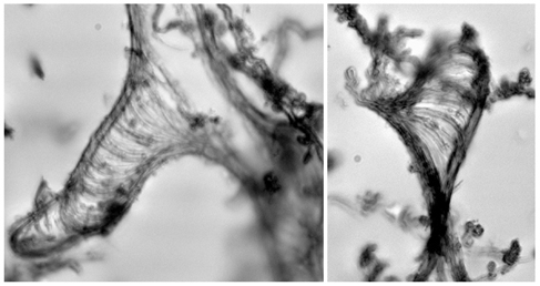
What are These?
There are unique structures within the mat that we cannot recognize as being directly associated with any of the bacteria present. Often they look like a piece of yarn or rope that is unraveling; perhaps that is analogous to what they are, iron fibers that are coming apart, or perhaps, they are formed by some other process or organism we don’t understand.
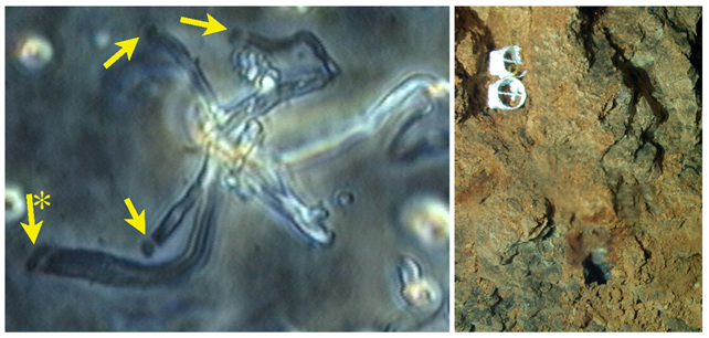
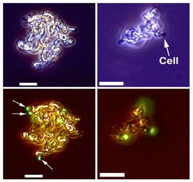
Where are the Cells?
When looking through the light microscope at the Loihi iron mats, one thing you quickly notice is that there are no cells visible. How can we say the bacteria are weaving the mats if we can’t see cells on the fibers? This puzzled us for some time, but we came up with a simple solution to see the cells attached to the iron oxides. This was done by putting special microscope slides down at the vents for short periods of time and bringing them back up and looking at them in the microscope. The image below is from one of these ‘slide traps’. The yellow arrows point to small bean-shaped cells that are growing at the end of the iron stalks or fibers. We now know that as the cells grow they extrude the stalk, in fact the stalk remains stationary and the cells move by extruding the stalk behind them. Perhaps this helps them both stay attached in an environment they like, and, at the same time, move from one place to another within that microenvironment. This also indicates that the attachment of the cell to the stalk is very delicate, the normal sampling and handling of samples probably causes most the cells that are on filaments to be broken off, which is why we normally do not see cells on the filaments.
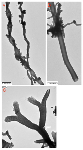
Fibers upon Fibers
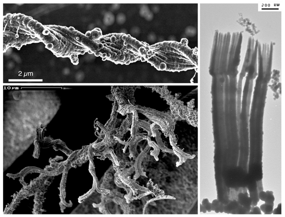
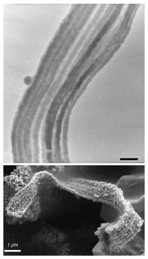
Perhaps even more remarkable is that by using a high-powered electron microscope we can observe that the iron thread that an individual bacterium produces is itself composed of a bundle of even finer iron threads. These fine filaments are the iron oxides that the cells excrete as they grow. The fine fibers are extremely thin, just a few nanometers across, hundreds of times thinner than a human hair. These bacteria are true nano-engineers. We know the fibers are made mostly of iron, but what else? We think the cells must be producing an organic molecule that makes the iron oxides form such uniform fibers, but we don’t know what it is. What keeps all the individual iron fibers stuck together?
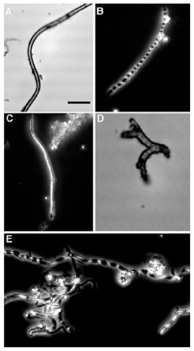
Mariprofundus ferrooxydans
This is an iron-spinning bacterium that we isolated from Loihi iron mats. It forms a twisted stalk. It was isolated by growing it in opposing gradients of ferrous Fe(II) iron, which is soluble, and oxgyen. The twisted stalk-like threads are one of the most common morphologies of iron oxides that we see in the fabric of Loihi microbial mats. We have also used molecular-based tools that do not require growing the organism to show that Mariprofundus-like bacteria are common at Loihi and in other iron mats associated with deep-sea vents. M. ferrooxydans is well suited to growing at Loihi, the only energy source it can use is ferrous iron, it basically has to eat nails for a living. By being able to study it in the lab we hope to learn more about its remarkable talent for producing threads of iron, as well as understand other aspects of its physiology and behavior that allow it to thrive under such harsh conditions.
Dave Emerson onboard the R/V Kilo Moana
| FeMO2 Cruise Home Page |